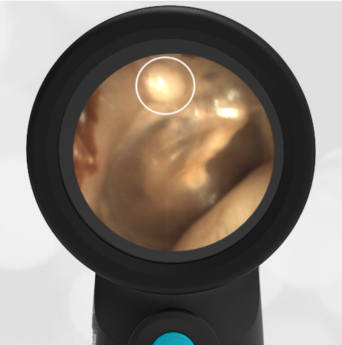
Osteoma
A 20-year-old female presents to the emergency department (ED) for a headache and sore throat. An incidental physical finding was discovered on her WiscMed Wispr digital otoscope exam. Upon further questioning, the patient denies being a surfer and does not enjoy cold water bathing or swimming.
Regarding the area circled on the image, which feature(s) help determine the most likely diagnosis:
- The lesion is solitary and unilateral
- The patient has never surfed nor had repeated exposure to cold water
- The lesion appears to have a slightly pedunculated base
- Histologic examination of the lesion does not demonstrate layers of new bone
- All the above

The incidental finding on this patient’s Wispr digital exam is most consistent with a small osteoma in the superior wall of the external auditory canal (EAC). An osteoma is a benign bony neoplasm that can be easily confused with an exostosis, the other common lesion found in the EAC. Osteomas are generally single, unilateral, and have a pedunculated base while exostoses are often multiple, bilateral, and have a wider base.

The two growths can be difficult to distinguish in mild cases by appearance alone. However, they are histologically distinct with exostoses having a laminated appearance representing layers of newly formed bone. This is believed to occur when repeated exposure to cold water triggers periostitis resulting in sequential episodes of osteogenesis, giving rise to the term “surfer’s ear.” Osteomas arise from cancellous bone and lack the layered growth pattern of exostoses. Both lesions are generally asymptomatic unless they enlarge enough to cause conductive hearing loss. In these instances, referral to otolaryngology (ENT) is warranted for further evaluation and potential surgical removal.
Reference:


