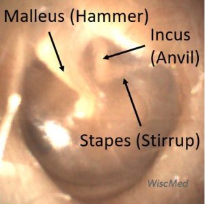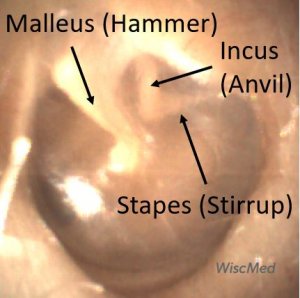
Hammer, Anvil & Stirrup by otoscopy. Bones of the middle ear.
There are three small bones (ossicles) that transfer the movement of the tympanic membrane (ear drum) caused by sound waves to the inner ear. The formal name of the three bones are malleus, incus and stapes. The more common names are hammer, anvil and stirrup. This chain of bones is an elegant example of natures bioengineering. It is small, efficient and reliable. When you view the bones using an otoscope, you are actually viewing them in the "middle ear." This is the space behind the tympanic membrane (ear drum). The reason that they can be seen is because the ear drum is somewhat transparent.

The bones of the middle ear are seen by the Wispr digital otoscope through a partially transparent tympanic membrane (ear drum)

The bones (ossicles) of the middle ear seen through the tympanic membrane (ear drum)
The malleus (commonly called the “hammer”) is the first bone in the chain of three that translate movement of the tympanic membrane to the inner ear. The malleus is the most prominent landmark visible in the middle ear space. The malleus is also an anatomical compass in that it “points” to the face. This allows you to determine that this is the left ear.

Malleus and its Components
The portions of the malleus that are visible on otoscopic examination include the umbo, manubrium, and the short process. The short process is often the last portion of the malleus that is discernable in cases of infection as in this example. The malleus is attached to the tympanic membrane (eardrum). As the tympanic membrane vibrates from sound waves, the malleus converts this vibration into a rotary motion at the head (not visible) that is connected to the incus and then to the stapes. 
Malleus, Incus and Stapes (Hammer, Anvil and Stirrup)
This is a beautiful example of a normal and healthy ear. Notable in this image is the ability to see all three bones of the middle ear through the transparent tympanic membrane; the malleus, incus, and stapes. It’s often not possible to see the stapes as it is the deepest bone in the middle ear space. The stapes connects the incus to the oval window of the inner ear, allowing the mechanical energy of the tympanic membrane to be communicated to the fluid in the inner ear. The malleus is almost always visible except in cases of distorted anatomy such as from acute otitis media. The incus is often visible, but normal differences in anatomy can hide this bone, too. All three bones of the middle ear are located in the posterior superior section of the tympanic membrane.
