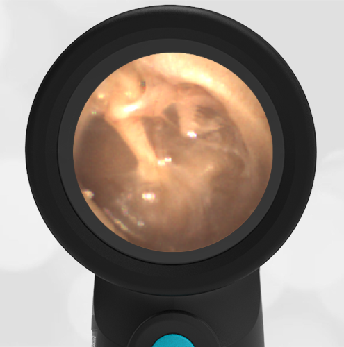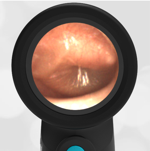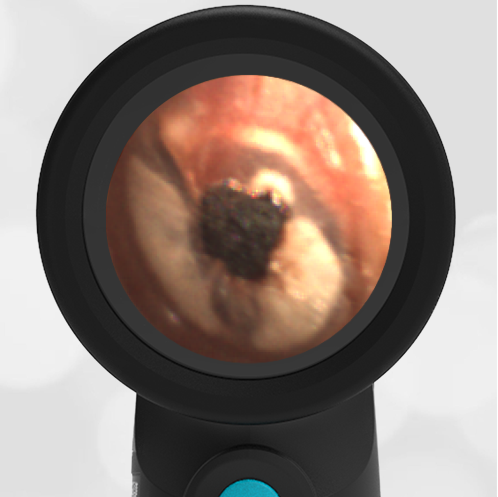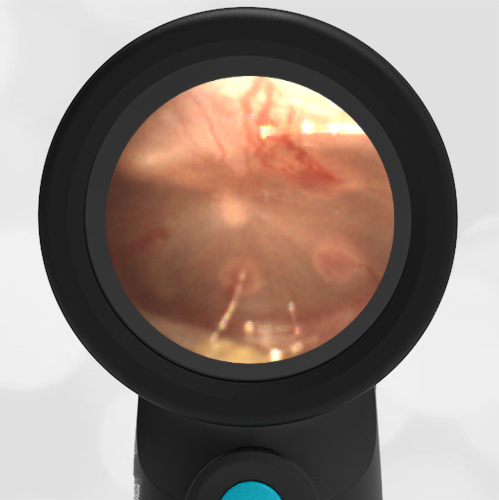
History of Tympanostomy Tubes – November 21, 2024
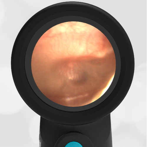
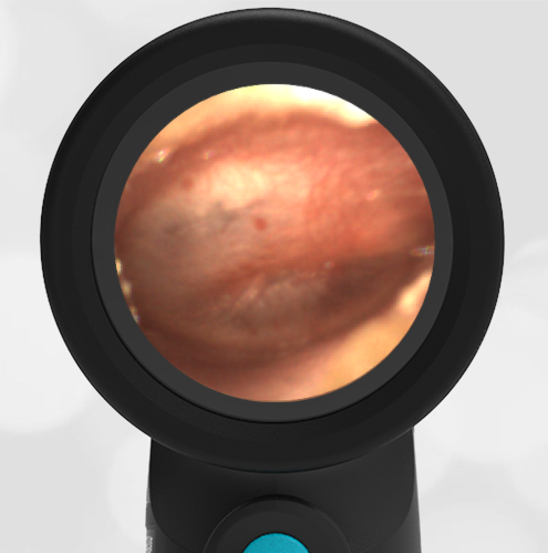
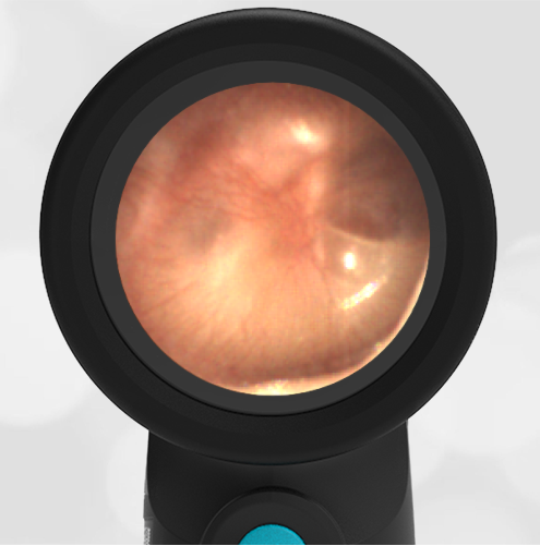
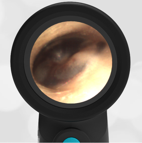
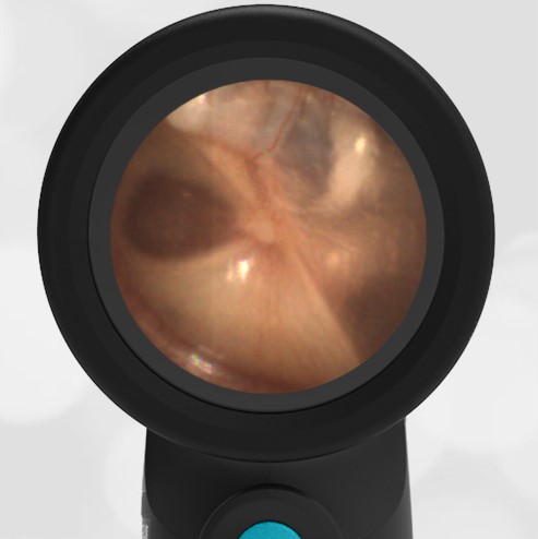
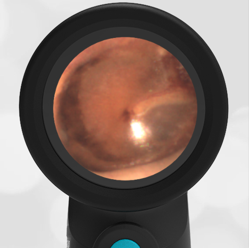
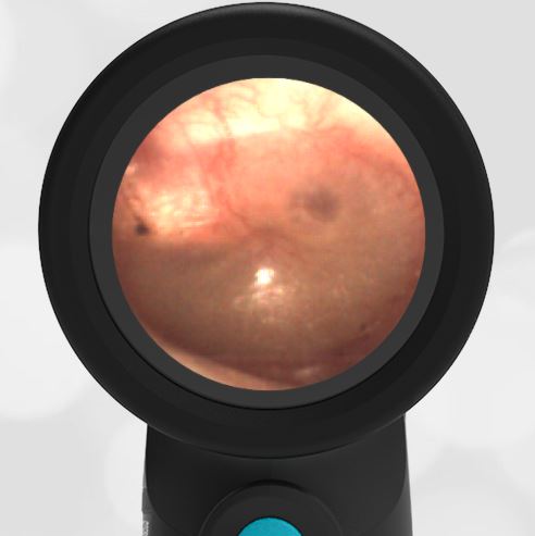
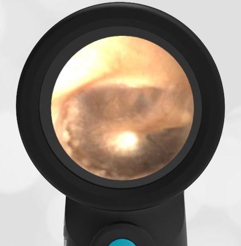
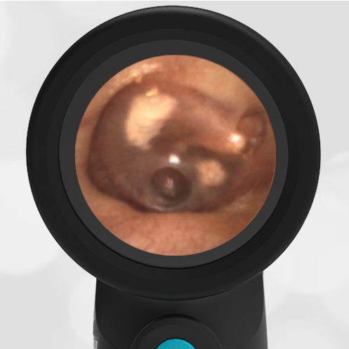
A 17-year-old male presents to the emergency department with foot pain. As part of a complete physical examination, this image of his right eardrum is obtained using the Wispr Digital Otoscope. He has no ear or hearing complaints. What caused the circular pattern seen in the inferior-anterior portion of the eardrum?
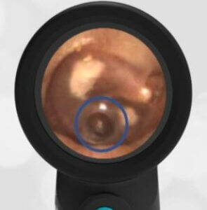
The circular defect in the ear drum (blue circled area) appears almost like a “punch biopsy.” It’s not clear what caused this pattern. An initial thought is that it is from a history of ventilation tubes (ear tubes, tympanostomy tubes). However, that seems unlikely as tube placement is accomplished by a linear incision in the ear drum which would not leave a circular pattern. From the presence of the sclerosis (white plaque), the patient has either had ear infections or ear tubes in the past.
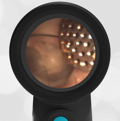
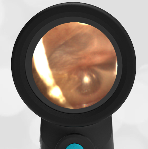
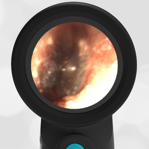
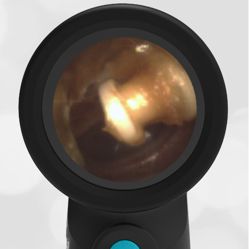
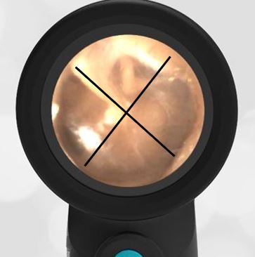
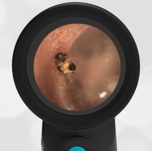
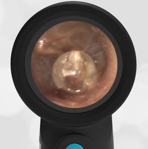
Ear tubes are often placed in children who have recurrent ear infections. Read our prior Interesting Image on tympanostomy tubes. The ear tubes eventually fall out. After the eardrum heals, there can be evidence of the prior placement (sclerosis), as in this image.
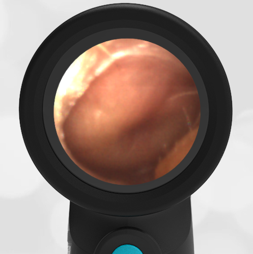
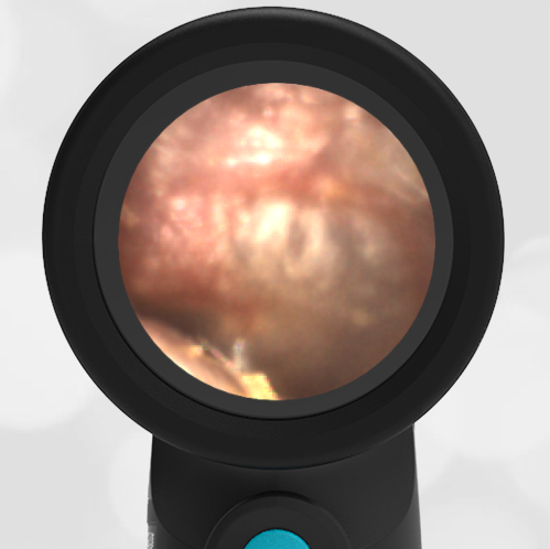
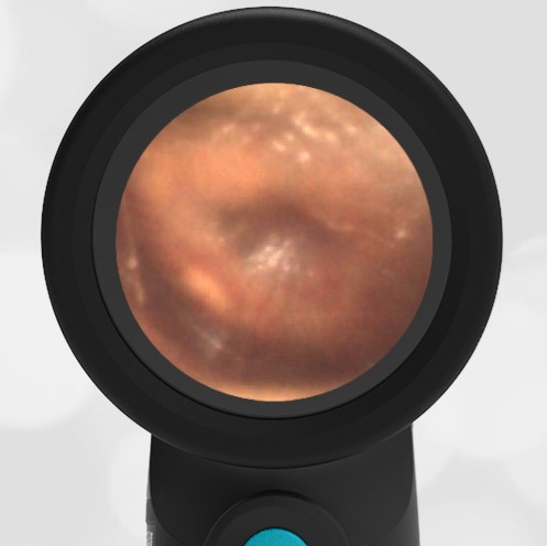
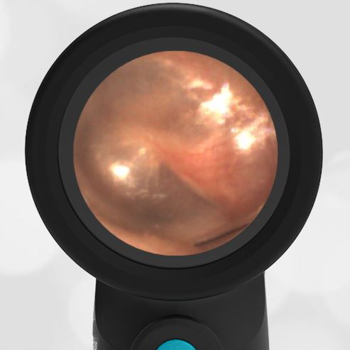
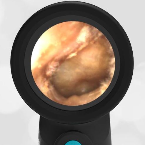
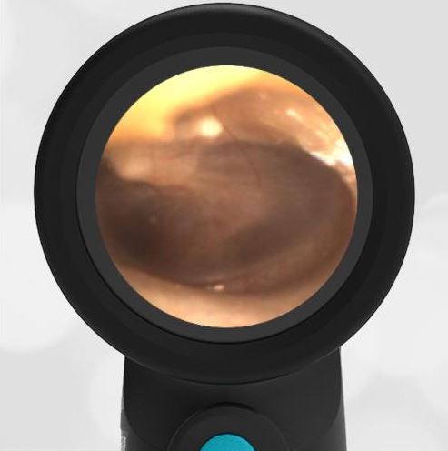
The patient’s left ear had a similar finding of post-tympanostomy tube healing and sclerosis as seen in this image:
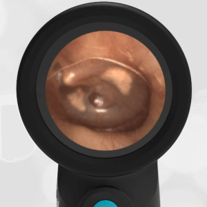
Patient’s left ear
Key learning points
- Tympanostomy (ear tubes) are commonly placed in children who have recurrent ear infections. The tubes drain effusion from the middle ear space and equalize pressure across the ear drum.
- The placement of ear tubes often leads to patches of sclerosis on the ear drum. These have little clinical significance.
- Tympanostomy tubes fall out on their own after about 6 months and the ear drum heals.


