Stapes Bone
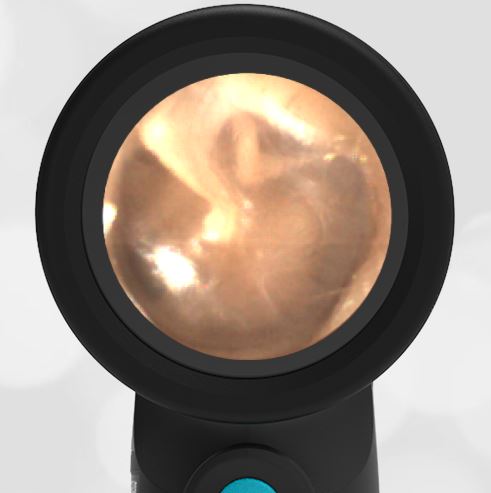
A healthy 30-year-old female had a demonstration of the Wispr digital otoscope at the 2021 American College of Emergency Physicians Scientific Assembly (ACEP). This image of her ear was obtained.
Can you identify all three bones of the middle ear?

This is a beautiful example of a normal and healthy ear. What’s particularly notable in this image is the ability to see all three bones of the middle ear through the transparent tympanic membrane, the malleus, incus, and stapes, commonly called the hammer, anvil, and stirrup. It’s usually not possible to see the stapes as it is the deepest bone in the middle ear space. The stapes connects the incus to the oval window of the inner ear, allowing the mechanical energy of the tympanic membrane to be communicated to the fluid in the inner ear. The malleus is almost always visible except in cases of distorted anatomy such as from acute otitis media. The incus is often visible, but normal differences in anatomy can hide this bone, too. More information on the anatomy and bones of the middle ear is available at Wispr University. Here is a video of this ear exam:


