Retraction – June 27, 2024
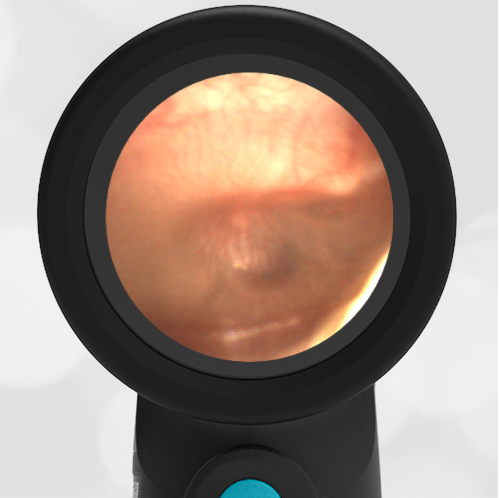
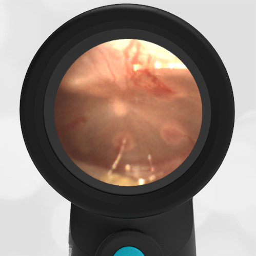
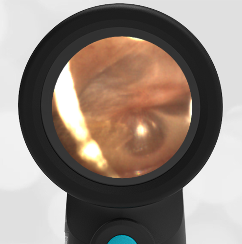
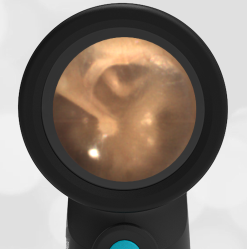
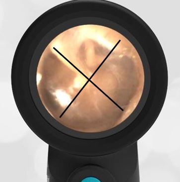
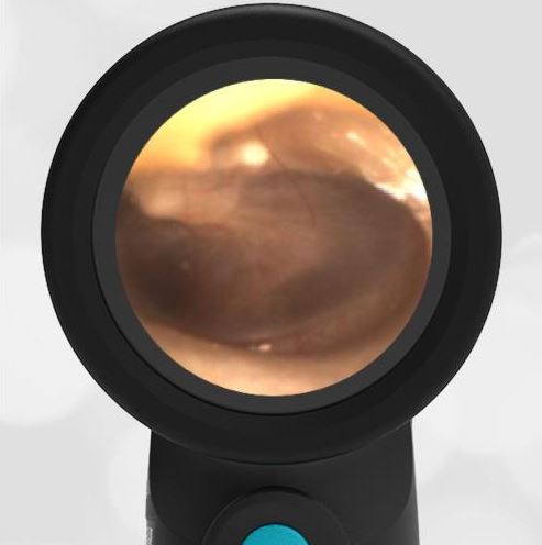
A 3-year-old female presented to the Pediatric emergency department (ED) with a complaint of congestion and ear pain. Her parents reported the child had multiple upper respiratory infections (URIs) since starting preschool and was treated for ear infections twice. Her most recent ear infection was approximately a month prior to the visit. Earlier in the evening, she was holding her hand over her ear—which her parents interpreted as indicating she was uncomfortable. The child had a runny nose for about a week; however, she has not had a fever. In the ED, she is well-appearing with normal vital signs and mild clear rhinorrhea. Her Wispr digital otoscopic exam is shown.
Which of the following is likely true regarding this child’s presentation?
- Her left tympanic membrane (TM) is retracted
- Her left TM is bulging
- Her right TM is retracted
- Her right TM is bulging
Answer: A. Her left tympanic membrane (TM) is retracted.

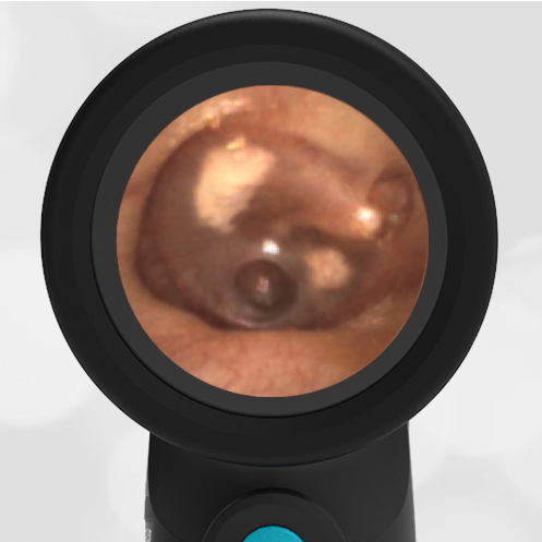
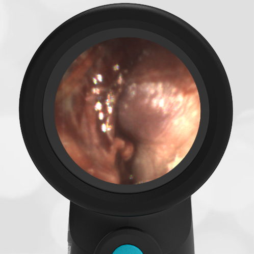
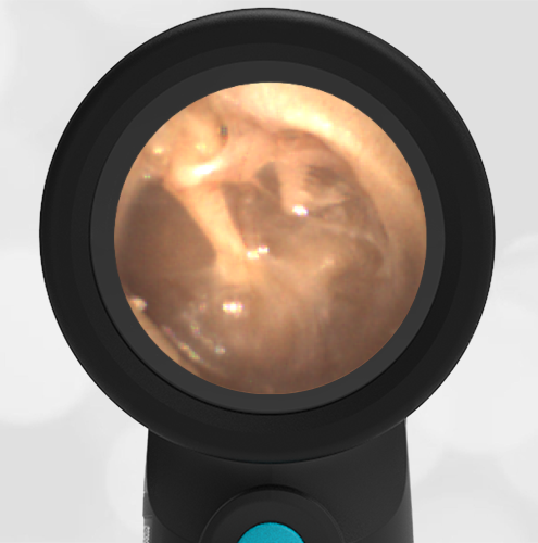
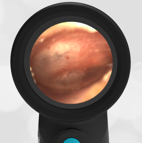
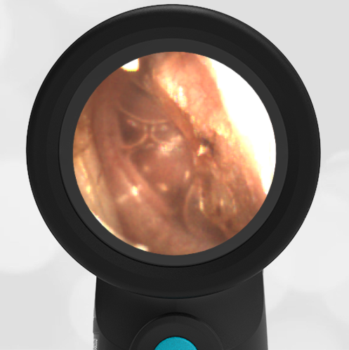
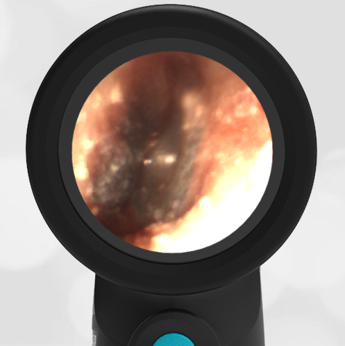
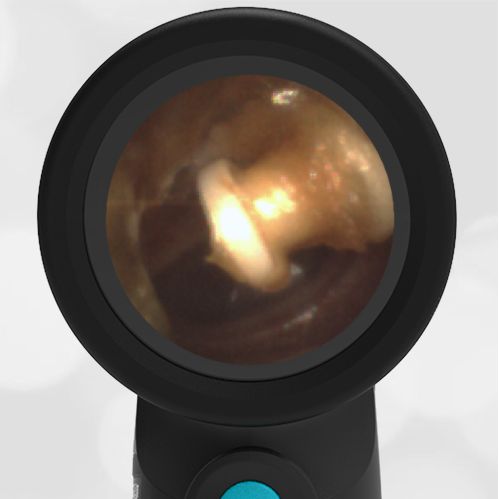
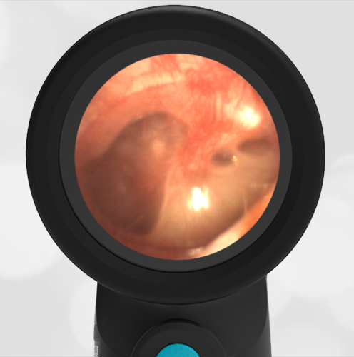
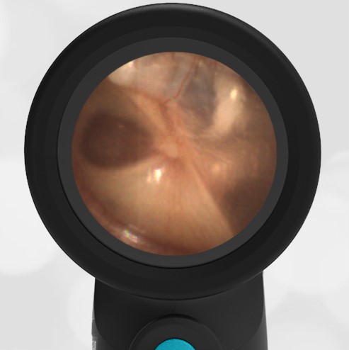
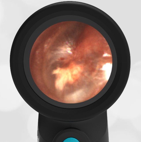
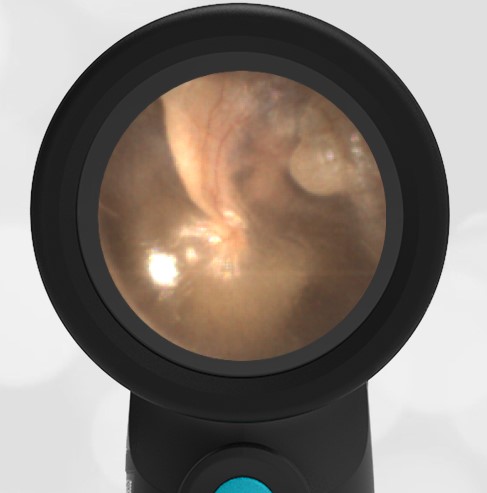
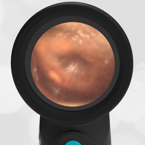
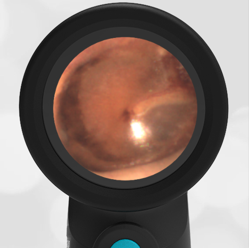
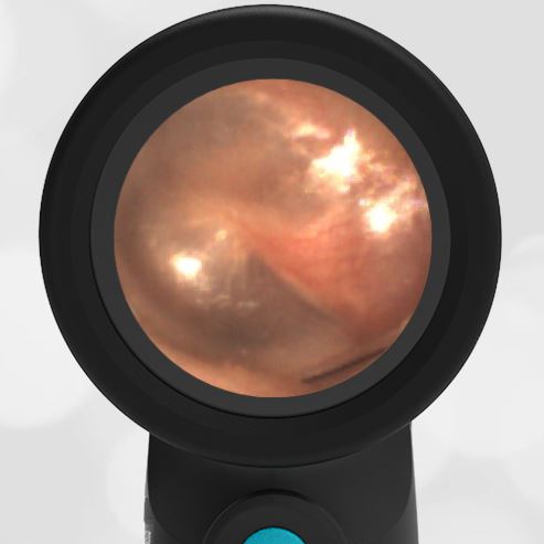
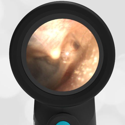
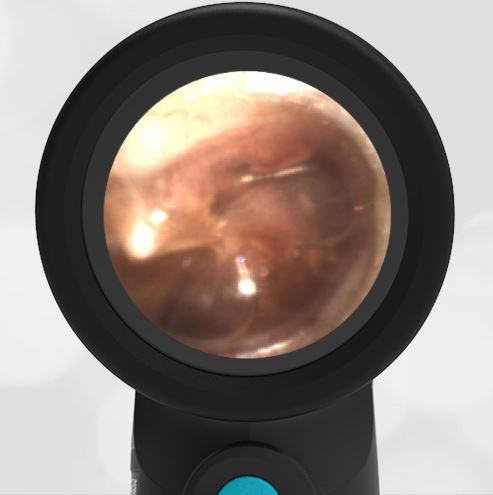
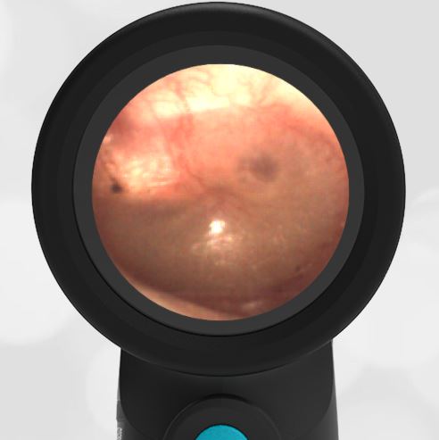
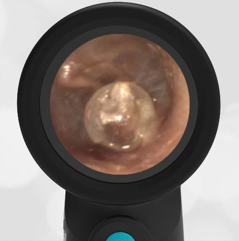
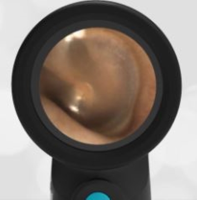
This child’s left TM is retracted as evidenced by the prominent lateral process of the malleus in the posterosuperior quadrant. While her retraction is mild, it is easily appreciated when compared to her right TM, which appears normal without the prominent lateral process. Ear laterally can be determined by the orientation of the malleus. Since the malleus is pointing up and to the left, this must be the left ear. Bulging such as from acute otitis media (AOM) is not present.
-

- Retracted Left TM
-

- Normal Right Ear
Retraction of the TM is often a sequela of eustachian tube dysfunction that blocks the normal pressure equalization between the middle and outer ear. As air is reabsorbed by the mucosal lining of the middle ear space, the negative pressure causes the TM to pull inward (i.e., retract) and may result in pain, muffled hearing, or ear popping. Due to children’s horizontally oriented eustachian tubes, frequent URIs, and adenoid hypertrophy, they are particularly prone to eustachian tube dysfunction. Transient TM retractions can be treated with supportive management and brief use of topical anesthetic if there is significant pain. An ENT referral is warranted if the retractions are persistent or worsening. Complications such as hearing loss, retraction pockets (deep localized retractions), progression into perforation, ossicular chain damage, or cholesteatoma may occur.
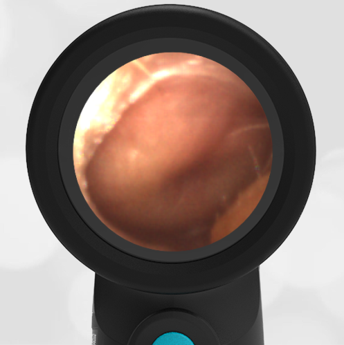
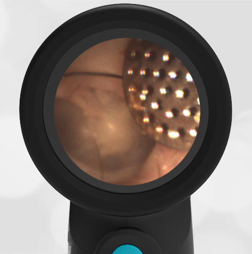
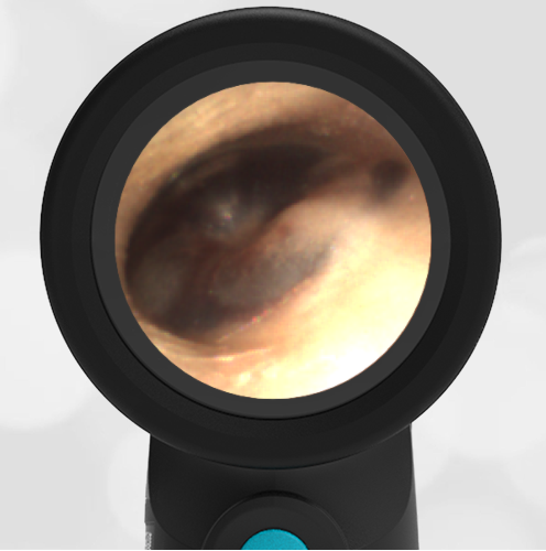
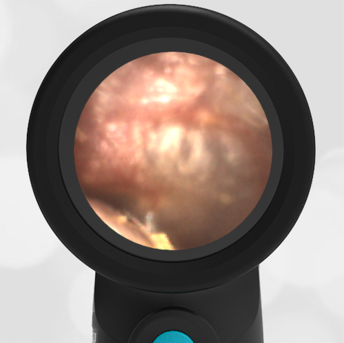
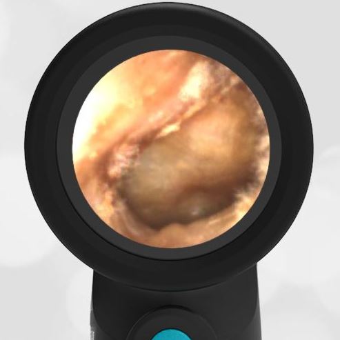
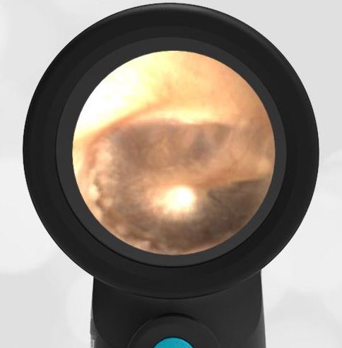
Here are the complete video exams of both the left and right ear.
Retracted Left TM Video
Normal Right Ear Video