TMJ Compression of External Ear Canal – October 3, 2024
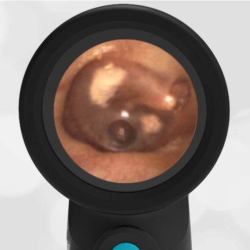
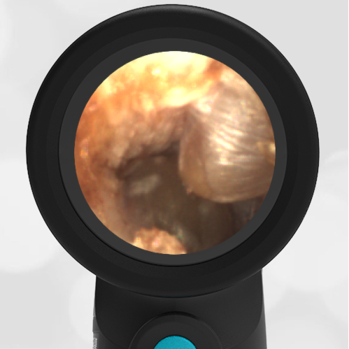
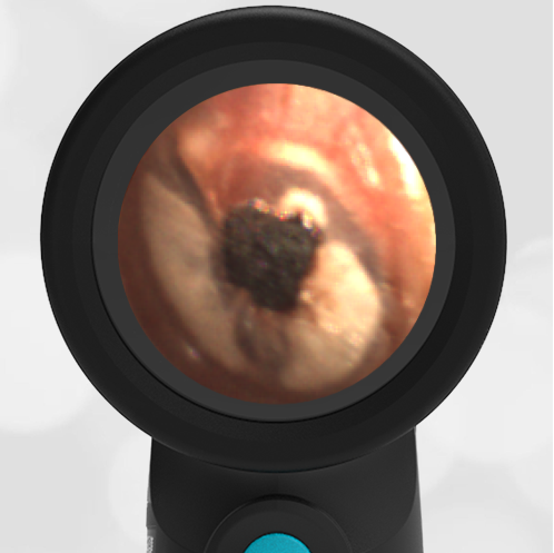
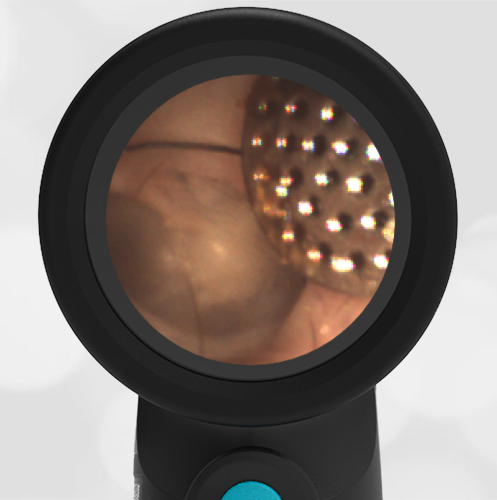
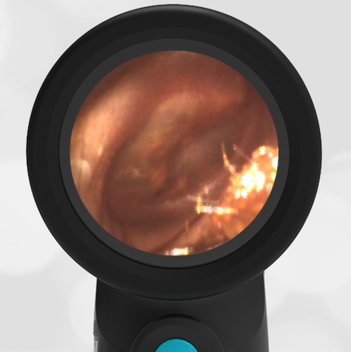
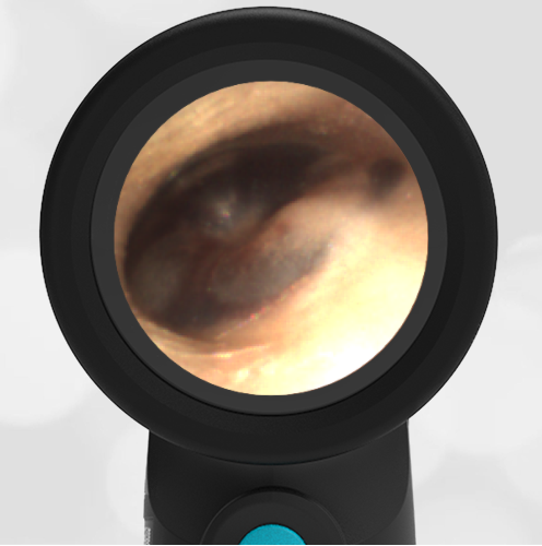
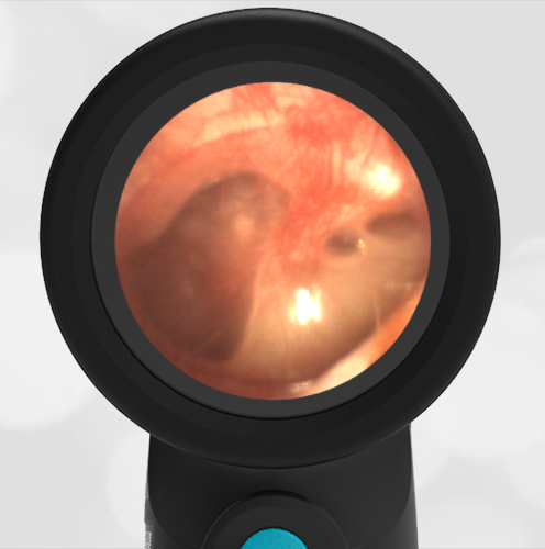
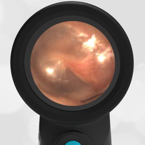
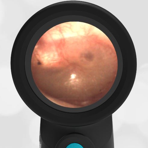
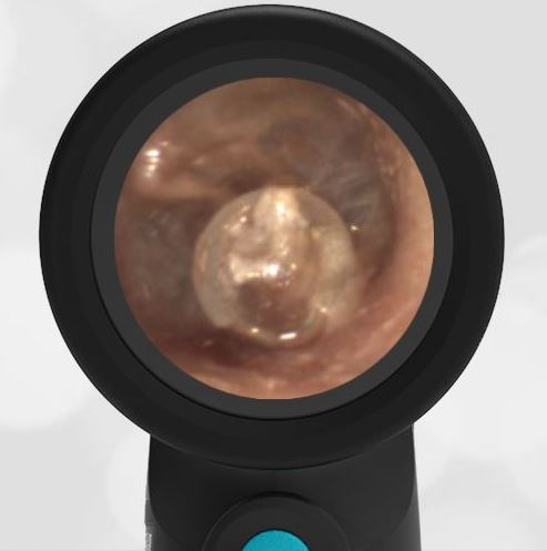
A 41-year-old woman was given a demonstration of the WiscMed Wispr digital otoscope at the American Academy of Pediatrics national convention. She mentions that in the past year, she has had increasing discomfort of her right temporomandibular joint (TMJ). She also says that the hearing in her right ear has significantly diminished. She has had no fevers, no unexplained weight loss, and no headaches. The following image of her right ear canal is obtained.
What is the next step in management?
- Urgent consultation to otolaryngology (ENT)
- Obtain a CT scan of the temporal region
- Begin antibiotics
- Begin a course of high-dose steroids
Answer 2. Obtain a CT scan of the temporal region

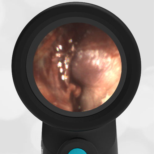
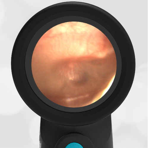
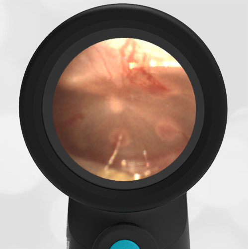
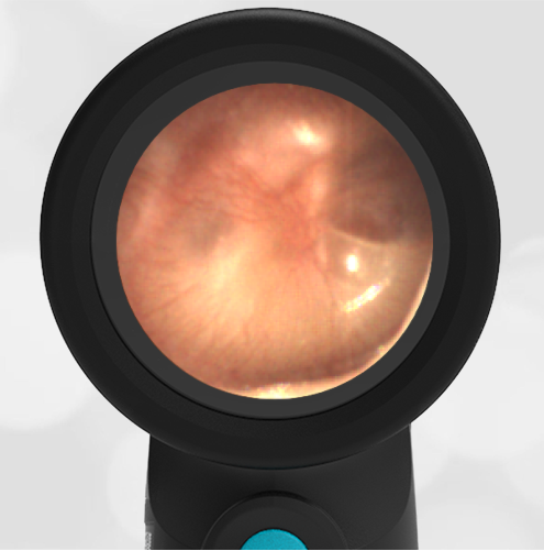
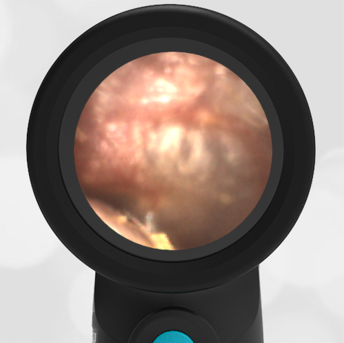
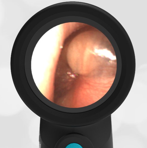
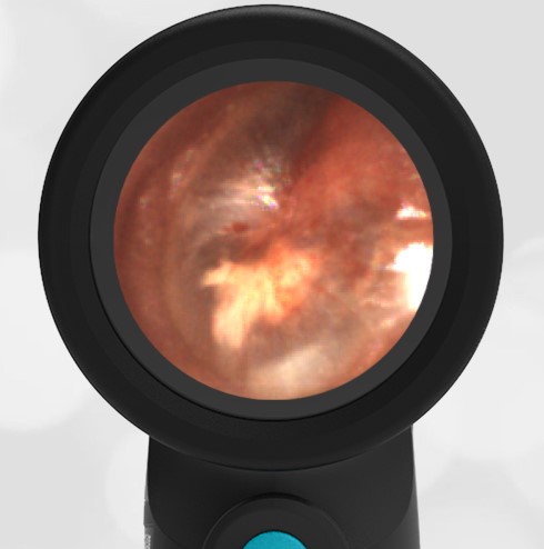
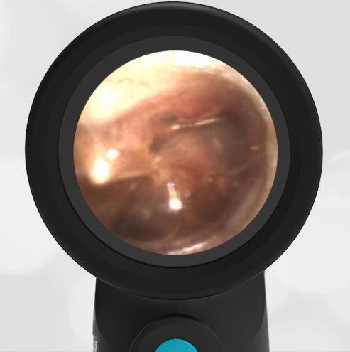
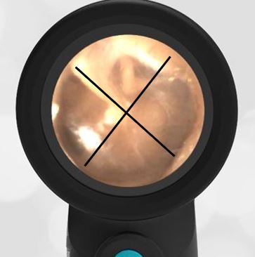
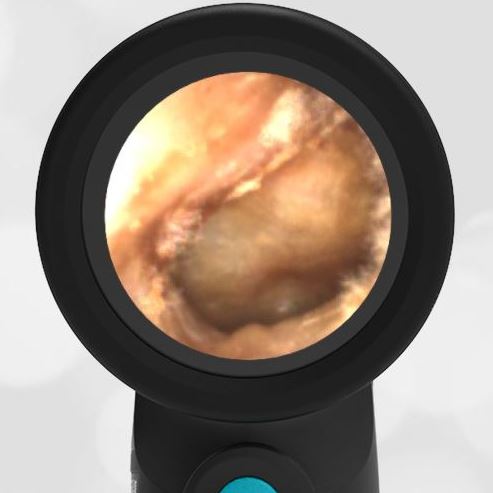
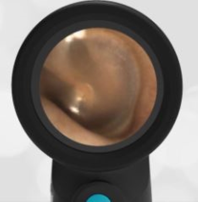
The woman has a significant occlusion of her external ear canal, likely caused by mechanical compression associated with the TMJ. Compare the image of this patient’s external canal with a normal canal. Note that a normal external canal is round while the patient’s canal is so compressed as to appear as a slit.
-

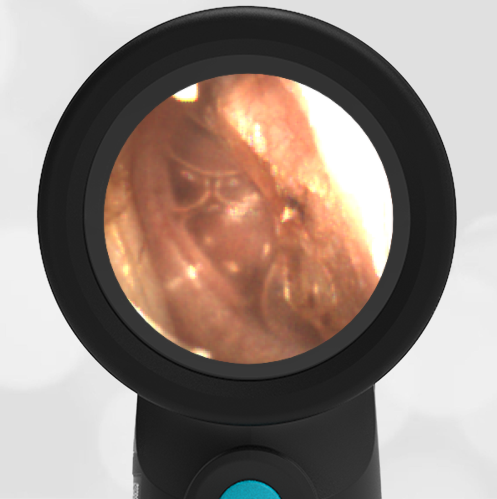
- TMJ Occlusion
-

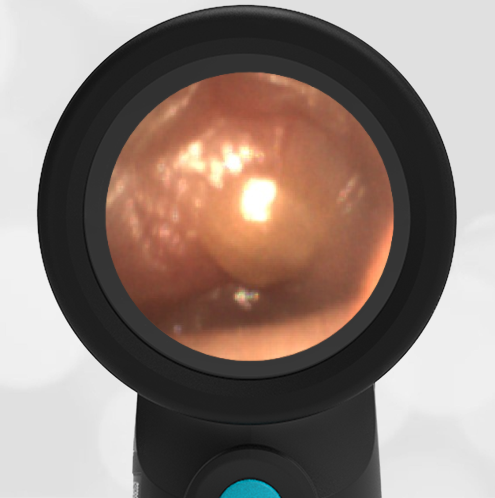
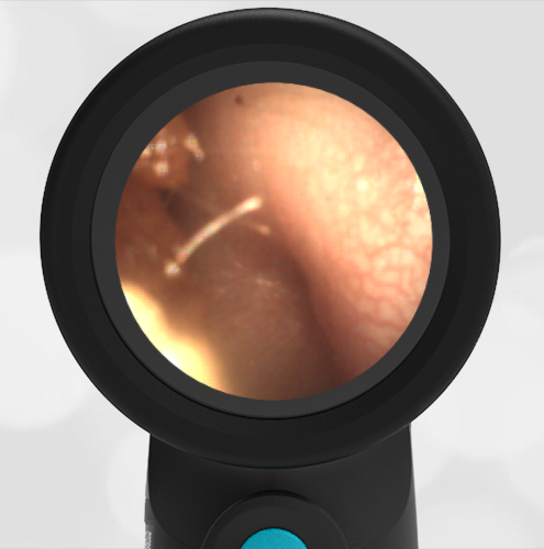
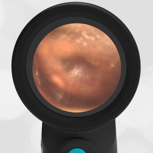
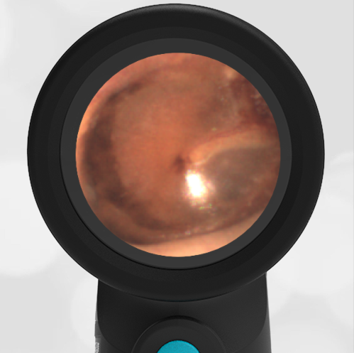
- Normal Right Ear
There is no indication of infection, i.e. no erythema or evidence of inflammation, so antibiotics are not indicated. High-dose steroids are not indicated until further information regarding the cause of the compression is determined. A CT scan or MRI would be the next step to identify the source of the compression and to eliminate the possibility of a tumor. After advanced imaging, a consult with otolaryngology would be appropriate.
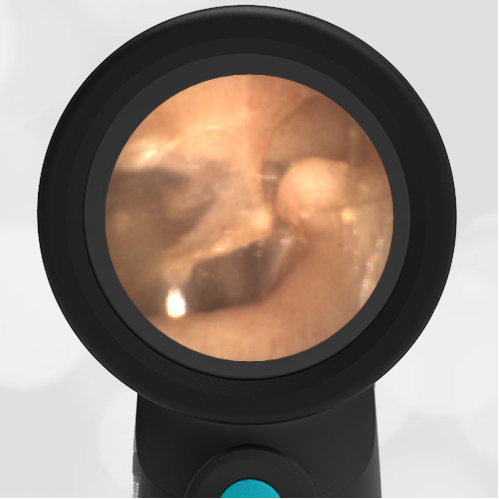
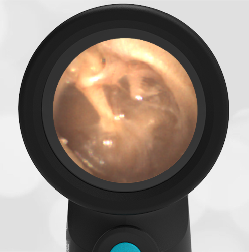
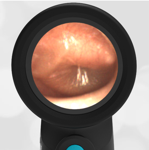
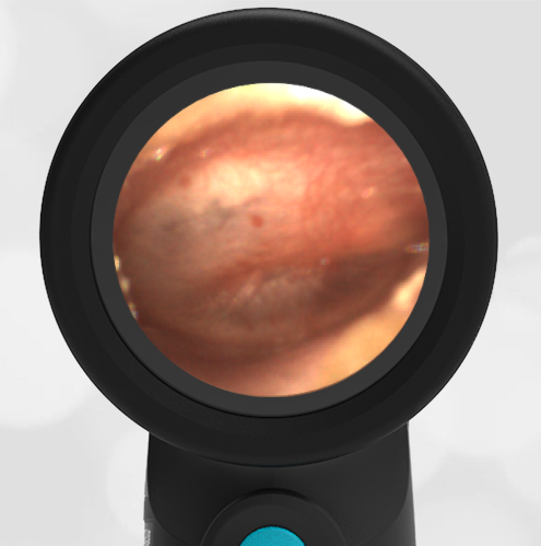
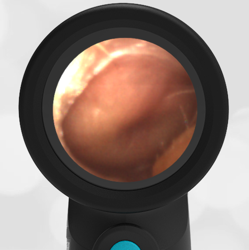
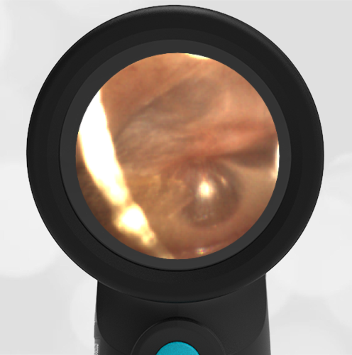
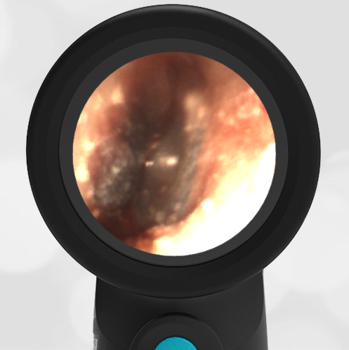
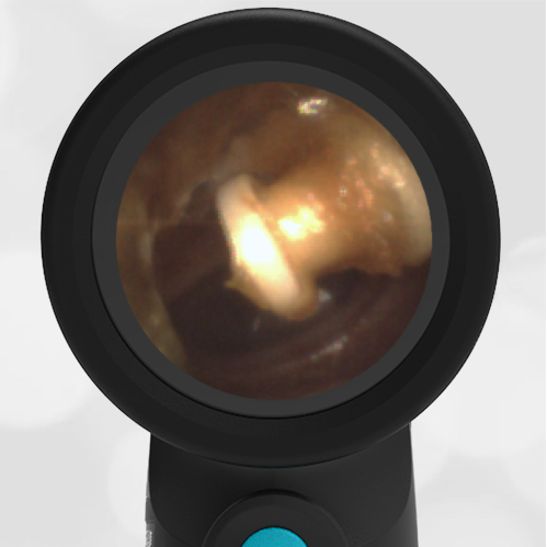
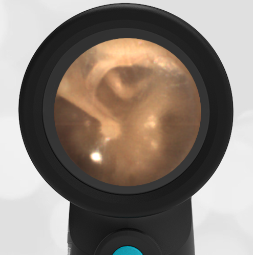
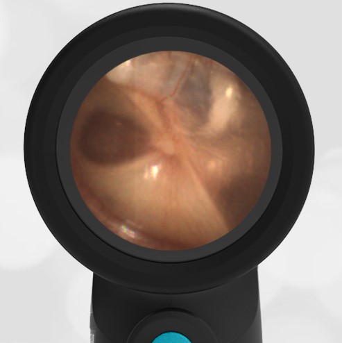
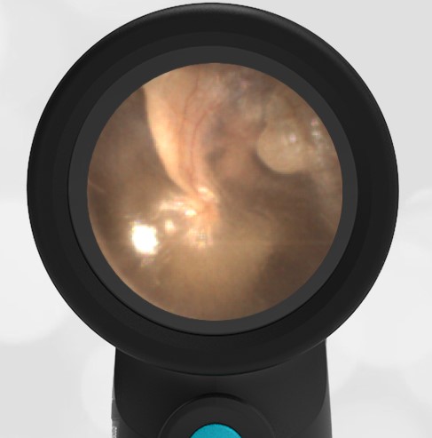
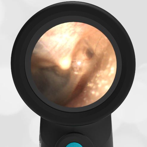
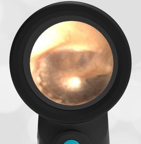
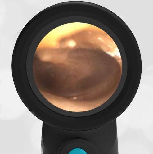
The patient was asked to open her mouth widely and move her lower jaw to the left. When she did so, the external canal opened slightly, and a partial view of the tympanic membrane (ear drum) was obtained. The visible portion of the eardrum appeared healthy and normal.

Here are the complete videos of the exams with both the mouth closed and the mouth open with the lower jaw displaced to the left.
Mouth Closed
Mouth open, lower jaw to the left