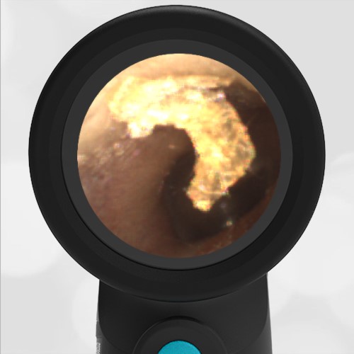
There are a number of choices available for digital otoscopes. We have previously reviewed 5 digital otoscopes here . While the basic components of each of these devices are the same – a digital camera, a light source and a display – how these components are implemented makes a vast difference in the clinical effectiveness of the device. A key design point for a digital otoscope is the location of the digital camera. The location of the camera determines the ability of the digital otoscope to obtain a diagnostic view of the tympanic membrane in the presence of obstructions such as cerumen.
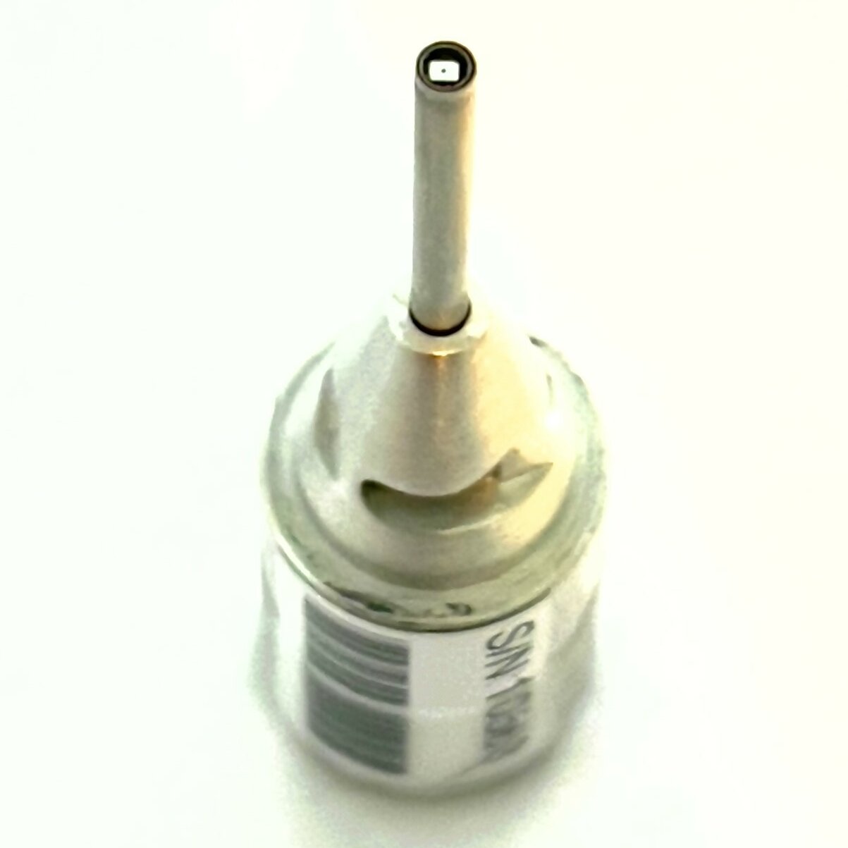
It is easiest to discuss the location of the digital camera relative to the speculum. The speculum being the disposable one-time use protection between patients. The camera may be located at either the “tip” of the speculum or the “base” of the speculum. The WiscMed Wispr digital otoscope is the only digital otoscope available with a camera at the tip of the speculum. You can see the Wispr camera at the end of the barrel in the image to the left. This location positions the camera at the tip of the speculum when the speculum is attached.
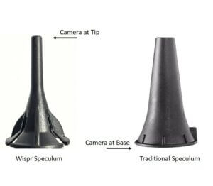
The advantage of having the camera located at the tip of the speculum is that it is possible to navigate the camera around obstructions in the ear canal to obtain a clear view of the tympanic membrane. This is not possible with either a traditional analog otoscope nor with a digital otoscope that has the camera at the base of the speculum. Another benefit of the camera mounted at the tip of the speculum is that the image never appears as a “tunnel.” It’s as if your eye is positioned right at the tympanic membrane.
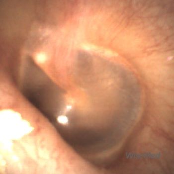
- Wispr image – non-tunneled
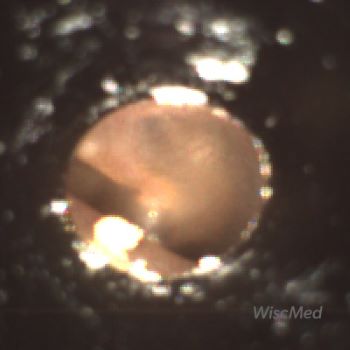
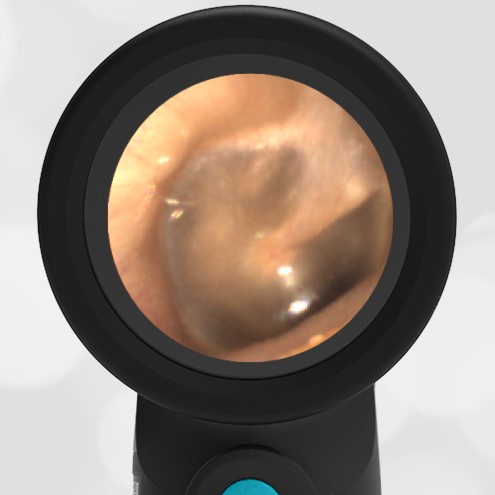
- Traditional image – tunneled
Here is an example demonstrating the benefit of the WiscMed Wispr’s camera placement at the tip of the speculum. This comparison demonstrates the view of the tympanic membrane that you might have with an analog otoscope to the view provided by the Wispr.
- Wispr digital view – past wax
- Traditional view – wax blocks ear drum
With the WiscMed Wispr and it’s digital camera at the tip of the speculum, it is easy to maneuver past the wax and obtain a diagnostic view of the tympanic membrane. This video demonstrates the exam on the same ear using the WiscMed Wispr. You can appreciate the navigation that allows for the clear view of the ear drum.
Video of clearing mild obstruction
You can clearly see the importance of camera location in reliably obtaining a diagnostic view of the tympanic membrane.

