Periosteal Abscess – May 2, 2024
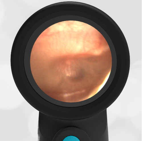
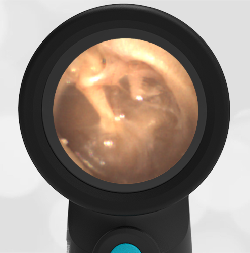
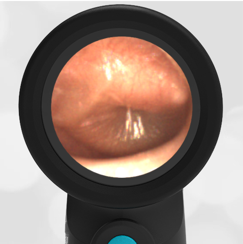
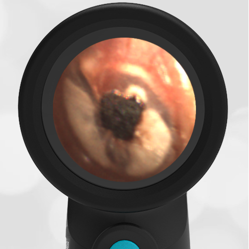
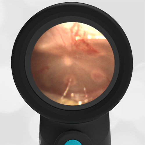
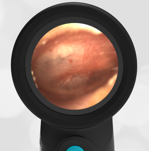
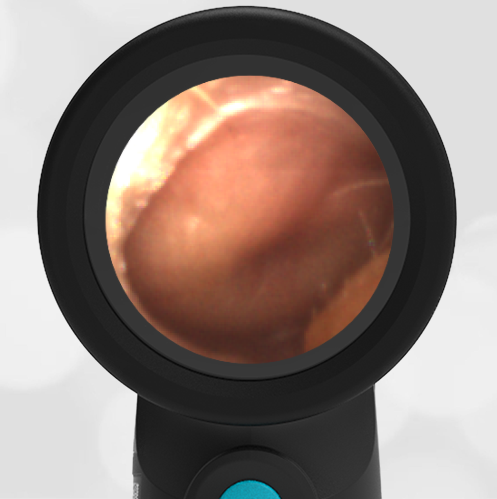
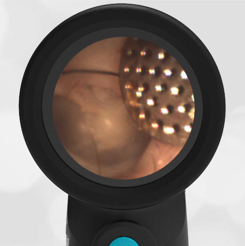
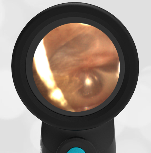
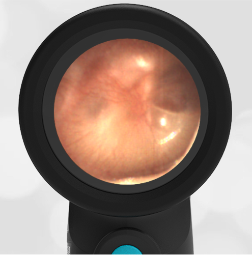
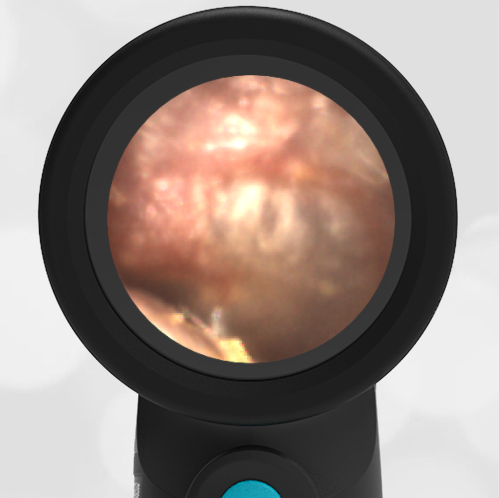
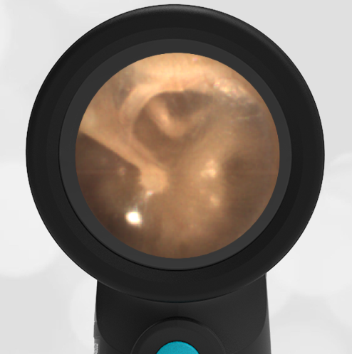
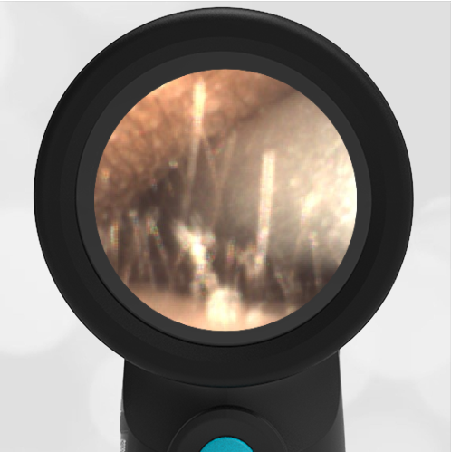
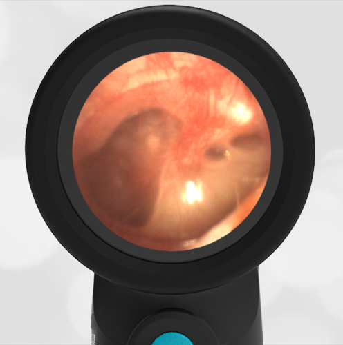
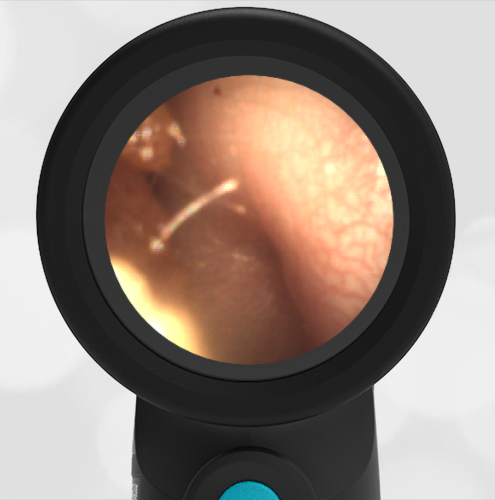
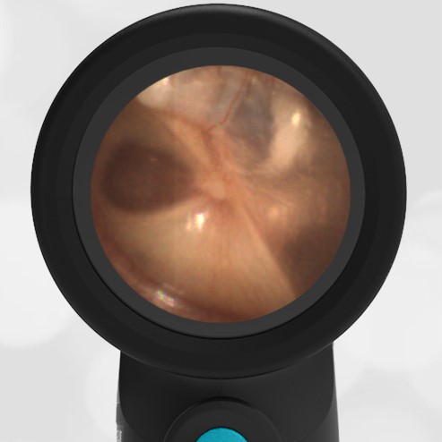
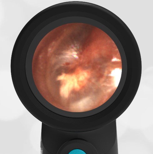
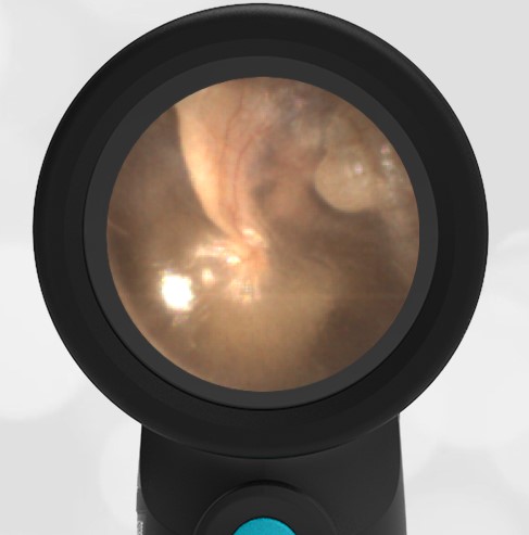
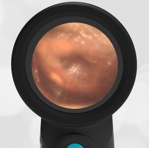
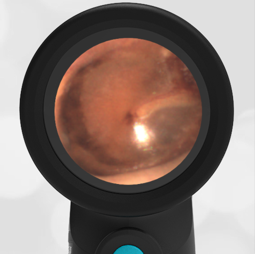
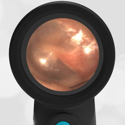
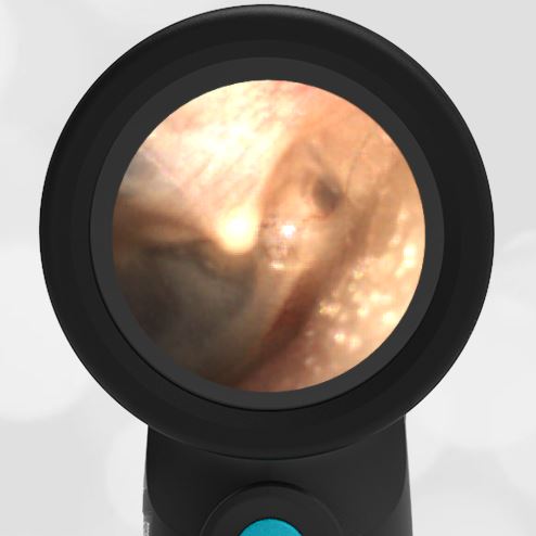
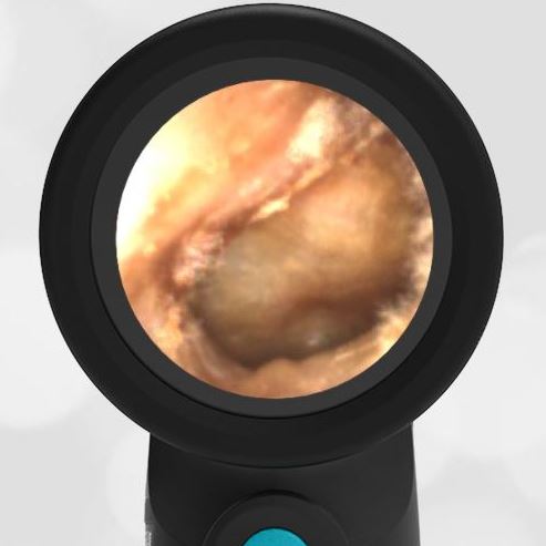
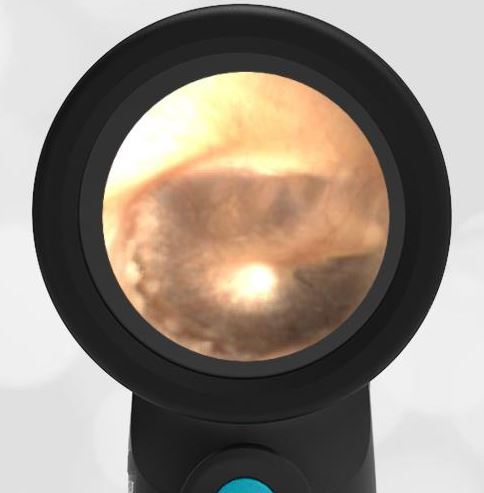
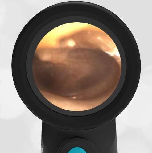
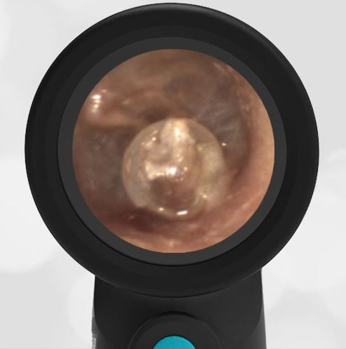
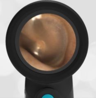
A 5-year-old female with mild autism is referred to the emergency department (ED) from Urgent Care (UC) for evaluation of an enlarged lymph node behind the right ear. The mother states the child does not complain of pain very often “even when sick.” She was concerned when her daughter had a fever of 103oF at home. At the UC, she was noted to have a tender lymph node which raised concern for an abscess. In the ED, the child was afebrile and generally well-appearing. Her HEENT exam was notable for her right external ear appearing quite prominent and a 3×3 cm semi-fluctuant posterior auricular mass with overlying skin erythema. Her left ear appeared normally positioned but with a smaller 1X1cm mass behind the external ear. Pulling externally on her ears did not elicit pain. Her right ear Wispr digital otoscope exam is shown.
Which of the following is true regarding the child’s presentation:
- She has acute otitis media (AOM) and should be treated with oral antibiotics and close follow-up.
- She has external otitis media (EOM) and should be treated with topical antibiotics and close follow-up.
- Imaging with contrast computed tomography (CT) is indicated.
- An ultrasound (US) of the posterior auricular mass with aspiration of any fluid for culture is indicated.
Answer: C. Imaging with contrast computed tomography (CT) is indicated.
The differential diagnosis of pediatric posterior auricular masses includes lymph node pathology, abscess, various cysts, hematoma, and occasionally malignancies. An ultrasound (US) is often helpful, and aspiration or incision and drainage may be necessary. However, in a child with fever, ear malposition, and posterior auricular swelling, cross-sectional imaging (CT) to evaluate for complications of mastoiditis such as subperiosteal abscess is indicated. The pathophysiology and complications of mastoiditis have previously been discussed here. Although the right external auditory canal does not appear normal, there is no significant swelling nor is there pain with manipulation of the external ear that would indicate external otitis media (EOM).

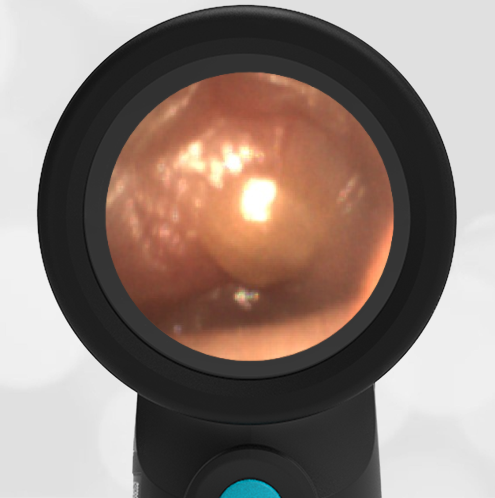
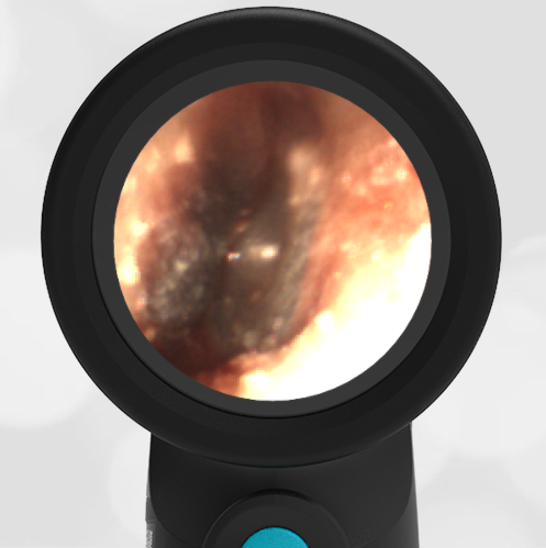
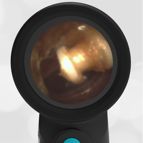
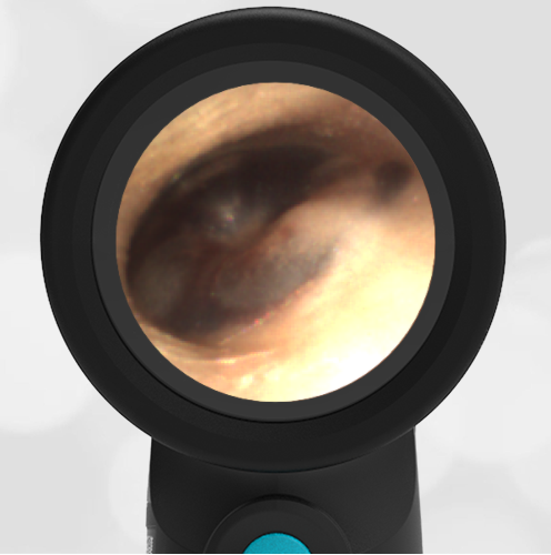
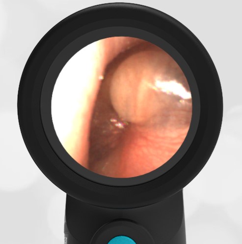
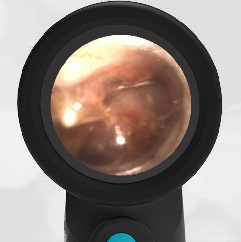
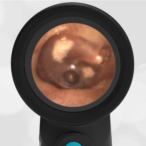
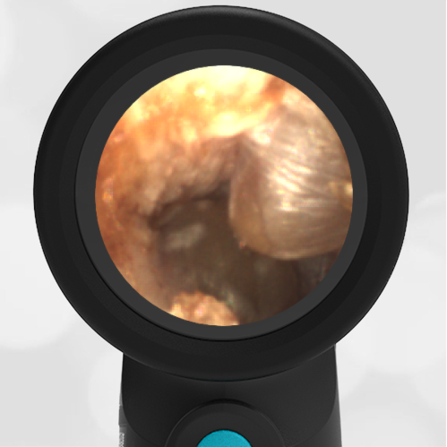
Looking carefully at the image, or the video (below), besides the obvious canal “blister,” the child also has acute otitis media (AOM) in the right ear. However, given the presence of the ear canal anomaly, concern is heightened for pathology more serious than simple AOM. The child had a contrast CT performed which demonstrated bilateral mastoiditis with subperiosteal abscesses. The blister-like appearing mass in the right external ear canal (EAC) was unroofed during surgery and noted to have serous fluid. The surgeon felt this was likely reactive from the nearby abscess.
Here is the complete video exam